2024 WINNERS
Morphological Differentiation of the F-Cell, a Specialized Epidermal Cell Embracing the Tactile Bristle
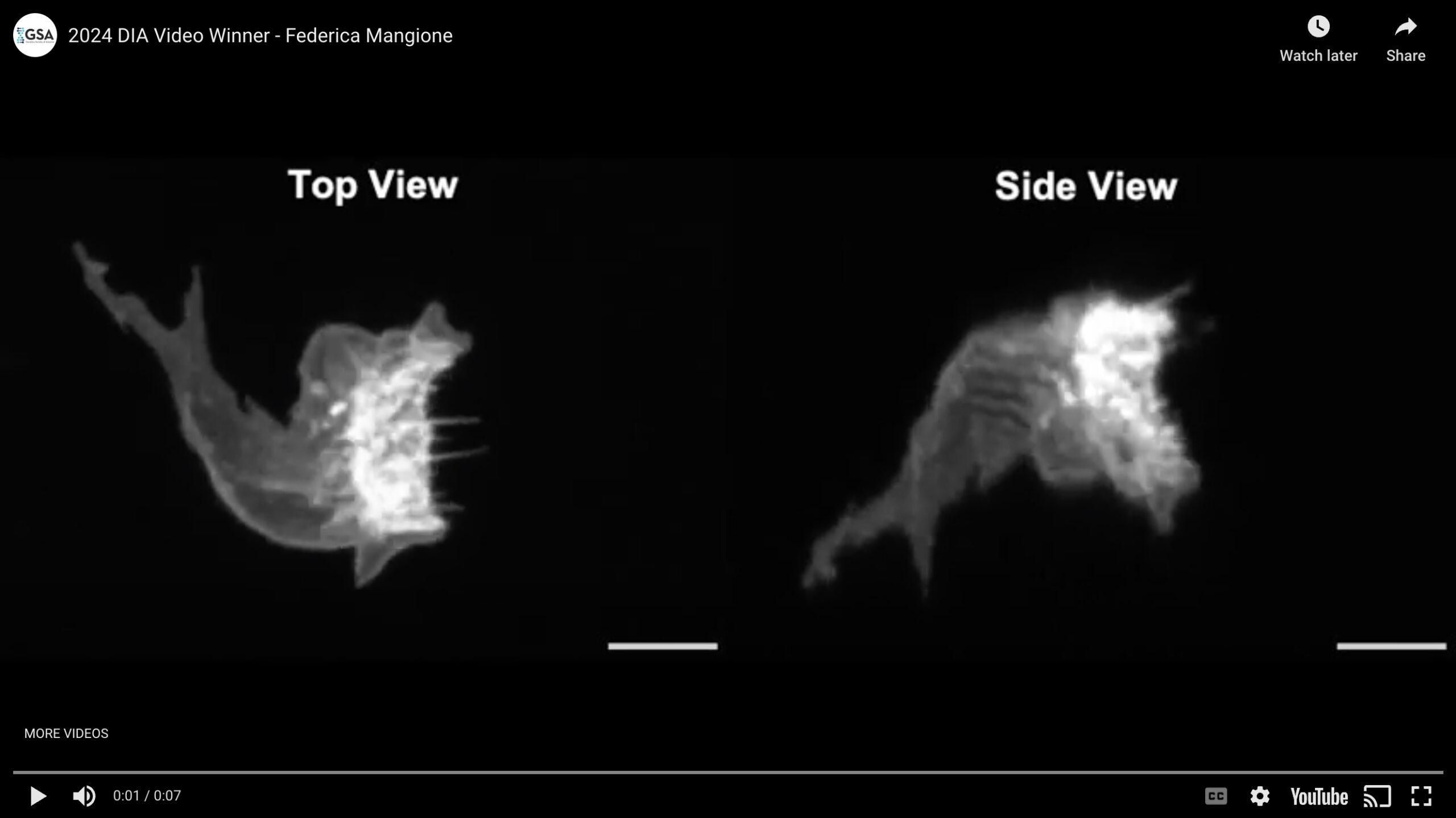
Specialized cellular structures endow the epidermis with the sense touch. While characterizing the terminal differentiation of the tactile bristles, the most abundant sensory organs within the Drosophila epidermis, we uncovered the association of a specialized epidermal cell, the F-Cell, by mid pupal stage. We found that F-Cells are recruited by the differentiating bristles from the surrounding epidermis to complete the assembly of the tactile bristle, enabling touch sensation in adulthood. The F-Cell and its differentiation were visualized for the first time in vivo by live imaging with confocal microscopy. This video captures the morphological changes of the F-Cell as it associates with a neighbouring bristle. Between 58 hours and 68 hours after puparium formation (hAPF), the morphology of the F-Cell changes markedly, culminating in a ring-like shape as it fully embraces the bristle by 68 hAPF. This unique shape distinguishes the F-Cell from other epidermal cells in Drosophila. For this video, the morphology of the F-Cell was made visible by the expression of the membrane marker myr::GFP under the control of 25c01-GAL4. Displayed scale bars are 5 µm. Our findings provide insight into the cellular organization underlying touch sensation and its developmental basis.
Video Credit: Federica Mangione
Mangione F, Titlow J, Maclachlan C, Gho M, Davis I, Collinson L, Tapon N.
Co-option of epidermal cells enables touch sensing.
Nat Cell Biol. 2023 Apr;25(4):540-549.
Cellular organisation of the fly’s kidney
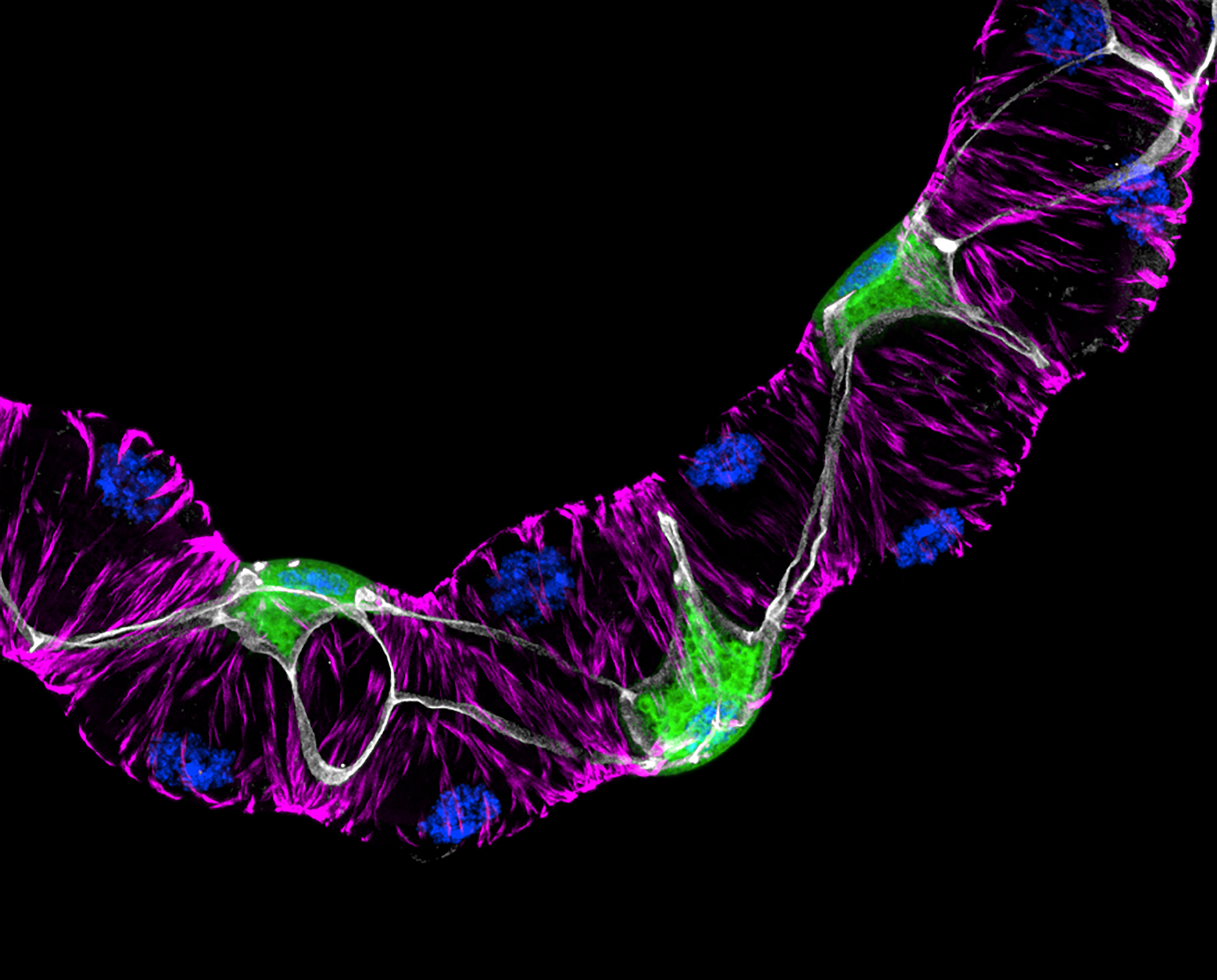
Detail of an adult Drosophila Malpighian (renal) tubule expressing membrane-bound GFP (green) in stellate cells – a population secondary intercalated cells required for transport of water – under the direction of c724GAL4, with staining of nuclei (DAPI, blue) and F-actin microfilaments (TRITC-conjugated phalloidin, magenta) in principal cells and stellate cells. Septate junctions are stained using an antibody for the junctional component Discs-large (anti-Dlg1, grey). The image was created by tiling of three separate maximum-intensity z projections obtained using a Nikon AX R NSPARC confocal.
Image Credit: Anthony Dornan
Dornan AJ, Halberg KV, Beuter LK, Davies SA, and JAT Dow.
Compromised junctional integrity phenocopies age-dependent renal dysfunction in Drosophila Snakeskin mutants.
J Cell Sci. 136(19) (2023).


